2027 webinars
- Webinar 1 - January 20
- Webinar 2 - February 17
- Webinar 3 - March 17
- Webinar 4 - April 14
- Webinar 5 - May 5
- Webinar 6 - June 16
- Webinar 7 - July 7
- Webinar 8 - September 22
- Webinar 9 - October 13
- Webinar 10 - November 17
- Webinar 11 - December 15
When RECIST Gets Difficult
Wednesday, January 20, 18:00 CET
Speaker: Giovanni Cappello, Turin/IT
Moderator: Evangelos Chartampilas, Thessaloniki/GR
General description, scope and objectives of this webinar
The programme of this webinar will cover challenging situations in tumour response assessment using RECIST criteria. It is aimed at for all radiologists, including those specialising in oncology, as well as residents and junior colleagues, given the clinical importance of this topic and who want to improve their skills in:
Applying RECIST criteria in complex and borderline clinical scenarios
Selecting and measuring target and non-target lesions consistently
Recognising common pitfalls that may lead to incorrect classification of treatment response
After watching the webinar, participants will be able to:
Apply RECIST criteria more consistently in challenging cases
Recognise situations in which apparent progression or response may be misleading
Provide clear and clinically relevant response assessments for multidisciplinary decision-making
Lung Cancer After Treatment: 5 Pitfalls That Can Change the Call
Wednesday, February 17, 18:00 CET
Speaker: Chiara Romei, Pisa/IT
Moderator: Giuseppe Cicchetti, Rome/IT
General description, scope and objectives of this webinar
The programme of this webinar will address five common diagnostic pitfalls encountered when assessing lung cancer after treatment. Case-based examples will illustrate how radiotherapy, systemic treatment and immunotherapy can produce imaging appearances that mimic residual or recurrent tumour.
It is aimed at for all radiologists, including those specialising in oncology, as well as residents and junior colleagues, given the clinical importance of this topic and who want to improve their skills in:
Recognising expected post-treatment appearances in lung cancer
Differentiating treatment-related changes from residual or recurrent disease
Integrating CT and PET/CT findings into response assessment
After watching the webinar, participants will be able to:
Identify key imaging patterns associated with different lung cancer treatments
Recognise common mimics of tumour progression and recurrence
Head & Neck Cancer After Treatment: Response, Recurrence or Treatment Effect?
Wednesday, March 17, 18:00 CET
Speaker: Aina Venkatasamy, Paris/FR
Moderator: Zuhir Bodalal (Elkarghali), Amsterdam/NL
General description, scope and objectives of this webinar
The programme of this webinar will focus on the challenging imaging assessment of head and neck cancer after treatment. Through case-based discussion, the session will explore how to differentiate expected treatment-related changes and complications from residual or recurrent tumour.
It is aimed at for all radiologists, including those specialising in oncology, as well as residents and junior colleagues, given the clinical importance of this topic and who want to improve their skills in:
Recognising expected post-treatment changes after surgery and radiotherapy
Differentiating treatment effect from residual or recurrent tumour
After watching the webinar, participants will be able to:
Avoid common pitfalls in post-treatment head and neck imaging
Provide structured reports that support appropriate further investigation and management
When Lugano Gets Difficult
Wednesday, April 14, 18:00 CEST
Speaker: Wolfgang Kunz, Munich/DE
Moderator: Lucien Beer, Cambridge/UK
General description, scope and objectives of this webinar
The programme of this webinar will focus on challenging aspects of lymphoma response assessment using the Lugano classification. Case-based examples will address situations in which PET/CT and morphological findings are difficult to interpret, with emphasis on avoiding common errors in response classification.
It is aimed at for all radiologists, including those specialising in oncology, as well as residents and junior colleagues, given the clinical importance of this topic and who want to improve their skills in:
Applying the Lugano classification and Deauville score in clinical practice
Interpreting residual masses and FDG uptake after treatment
After watching the webinar, participants will be able to:
Distinguish likely viable lymphoma from common post-treatment mimics
Communicate response assessment clearly and effectively to the multidisciplinary team
Solid Ovarian Tumours: Malignancy, Mimics and the Grey Zone
Wednesday, May 5, 18:00 CEST
Speaker: Laure Fournier, Paris/FR
Moderator: Gayané Sargsyan, Yerevan/AM
General description, scope and objectives of this webinar
The programme of this webinar will focus on the imaging assessment of solid ovarian tumours, with particular emphasis on distinguishing malignant lesions from benign mimics and indeterminate findings. Case-based examples will highlight key morphological and functional imaging features that can help refine differential diagnosis and guide clinical management.
It is aimed at for all radiologists, including those specialising in oncology, as well as residents and junior colleagues, given the clinical importance of this topic and who want to improve their skills in:
Recognising the imaging spectrum of solid ovarian tumours
Differentiating malignant lesions from common benign mimics
After watching the webinar, participants will be able to:
Narrow the differential diagnosis in challenging or atypical cases
Provide clinically relevant imaging assessments that support multidisciplinary management.
Extra-Axial Masses: From Differential Diagnosis and Surveillance to Post-Treatment Follow-Up
Wednesday, June 16, 18:00 CEST
Speaker: Simona Gaudino, Rome/IT
Moderator: Irina Trofimenko, Lisbon/PT
General description, scope and objectives of this webinar
The programme of this webinar will focus on the imaging assessment of extra-axial intracranial masses across the clinical pathway, from initial differential diagnosis and surveillance to post-treatment follow-up. Case-based examples will address common and uncommon mimics, imaging features relevant to management, and the challenges of distinguishing treatment-related change from residual or recurrent disease.
It is aimed at for all radiologists, including those specialising in oncology, as well as residents and junior colleagues, given the clinical importance of this topic and who want to improve their skills in:
• Developing a structured differential diagnosis for extra-axial masses
• Identifying imaging features relevant to surveillance and treatment planning
After watching the webinar, participants will be able to:
• Identify imaging findings that influence follow-up and management
• Distinguish post-treatment change from residual or recurrent disease
After Pancreatic Surgery: Normal, Complication or Recurrence?
Wednesday, July 7, 18:00 CEST
Speaker: Chandan Kakkar, Ludhiana/IN
Moderator: Alessandra Borgheresi, Ancona/IT
General description, scope and objectives of this webinar
The programme of this webinar will focus on imaging after pancreatic surgery, with emphasis on differentiating expected postoperative appearances from complications and tumour recurrence. Through case-based examples, the session will review altered postoperative anatomy, common diagnostic pitfalls, and imaging findings that require further clinical attention.
It is aimed at for all radiologists, including those specialising in oncology, as well as residents and junior colleagues, given the clinical importance of this topic and who want to improve their skills in:
Identifying early and late complications after pancreatic surgery
Differentiating postoperative change from recurrent malignancy
After watching the webinar, participants will be able to:
Recognise expected imaging appearances following pancreatic surgery
Identify key imaging features of common postoperative complications
Distinguish suspicious recurrent disease from benign postoperative changes
Nodal Staging with Molecular Imaging: Strengths, Caveats and Discordant Cases
Wednesday, September 22, 18:00 CEST
Speaker: Clemens Cyran, Munich/DE
Moderator: Giovanni Cappello, Turin/IT
General description, scope and objectives of this webinar
The programme of this webinar will focus on the role of molecular imaging in nodal staging of cancer, with particular emphasis on its strengths, limitations, and discordant findings. Case-based examples will illustrate situations in which molecular and morphological imaging findings do not agree and how these discrepancies can influence staging and management.
It is aimed at for all radiologists, including those specialising in oncology, as well as residents and junior colleagues, given the clinical importance of this topic and who want to improve their skills in:
Understanding the strengths and limitations of molecular imaging for nodal staging
Interpreting discordant findings between molecular and anatomical imaging
After watching the webinar, participants will be able to:
Recognise imaging patterns that may lead to incorrect nodal classification
Integrate molecular and anatomical imaging findings into clinically relevant staging assessments
Prostate Cancer Staging: 5 Imaging Pitfalls That Change Management
Wednesday, October 13, 18:00 CEST
Speaker: Andrea Ponsiglione, Naples/IT
Moderator: Claudio Fanni, Pisa/IT
General description, scope and objectives of this webinar
The programme of this webinar will address five important imaging pitfalls in prostate cancer staging that may alter treatment planning. Through case-based examples, the session will focus on challenging findings related to local tumour extent, nodal disease, and metastatic spread.
It is aimed at for all radiologists, including those specialising in oncology, as well as residents and junior colleagues, given the clinical importance of this topic and who want to improve their skills in:
Assessing local tumour extent and features relevant to prostate cancer staging
Recognising pitfalls in extra-capsular extension and invasion of seminal vesicles
After watching the webinar, participants will be able to:
Recognise common causes of under- and overstaging in prostate cancer
Interpret key imaging findings that influence local and systemic staging
Head & Neck Cancer Staging: 5 Imaging Pitfalls That Change Management
Wednesday, November 17, 18:00 CET
Speaker: Caterina Giannitto, Milan/IT
Moderator: Simona Gaudino, Rome/IT
General description, scope and objectives of this webinar
The programme of this webinar will focus on five key imaging pitfalls in head and neck cancer staging that can significantly affect treatment decisions. Case-based examples will address challenging findings at the primary tumour site, patterns of local extension, and nodal disease.
It is aimed at for all radiologists, including those specialising in oncology, as well as residents and junior colleagues, given the clinical importance of this topic and who want to improve their skills in:
Assessing primary tumour extent and anatomical structures relevant to staging
Recognising challenging patterns of nodal metastatic disease
After watching the webinar, participants will be able to:
Identify key imaging features that influence T and N classification
Provide accurate and clinically relevant staging assessments for multidisciplinary decision-making
The ESOI Imaging Quiz (no CME)
Wednesday, December 15, 18:00 CET
Upcoming webinar
Christmas Case Series
Wednesday, December 16, 18:00 CET
No CME points
Open to everyone!
Participants will have an opportunity to win prizes!
Who can register for this webinar and how?
This webinar is offered ONLY to all ESOI members in good standing. Please find all information about ESOI membership here: ESOI Membership
Should you be in doubt of your current membership status with ESOI, please contact the Office at office@esoi.society.org.
Learn about the state-of-the-art in oncological imaging all from the comfort of your home or workplace!
General Information
Since 2015, ESOI has been proudly offering high quality webinars given by world class speakers on various topics in oncologic imaging. Our live webinars have been very well received by our members with consistently high turnout and engagement from participants all across the globe. After the presentation, you can even ask questions in real time!
ESOI webinars are oriented towards different levels of expertise, a wide array of topics, and are increasingly becoming more multidisciplinary.
A number of our webinars are carried out in collaboration with medical societies as well as other radiological subspecialty societies.
Young radiologists are also included in the webinar series with special sessions addressing relevant topics for residents.
In addition to the live broadcast, the lectures are uploaded to the Member Area (with an approximate one month delay) so they can be (re)viewed by active ESOI members at their own convenience.
To date, we have more than 90 full length lectures (with in-depth discussions and Q&A)!
Recommended Bandwidth
The website www.speedtest.net allows you to check your internet bandwidth. We strongly recommend the following minimum requirements:
• Upload min. 6Mbit required bandwidth (especially important for speakers and moderators who share their video and audio signals)
• Download min. 6Mbit required bandwidth
For optimal stability, we recommend using a wired internet connection or a wireless connection with full signal strength.
Please note: Shared networks (e.g., in hospitals or universities) may significantly reduce the available bandwidth for each user.
Please Note
Webinar registrations are issued to an individual person and may neither be shared nor transferred to others under any circumstances.
The use of automated systems, AI tools, or any form of proxy attendance in place of the actual attendee is strictly prohibited.
Such actions are considered a violation of our participation policy and may lead to consequences, including revocation of the Certificate of Attendance, denial of CME credits and/or exclusion from future ESOI educational activities. We appreciate your understanding and cooperation in maintaining the integrity of our educational offerings.
Exclusive Sponsor 2026

Webinar Accreditation 2026
We are pleased to inform you that all regular webinars 2026 have been granted 1 European CME credit (ECMEC®s) by the European Accreditation Council for Continuing Medical Education (EACCME®).
Attendees need to attend the live webinar AND evaluate the webinar within a week to receive the CME certificate.
The certificate of attendance/CME certificate will be sent approx. 1 week after the live webinar.
Webinar Overview 2026
Check out the webinar schedule 2026 and mark your calendar!
Webinar Schedule 2026
Webinar 1
Tuesday, January 13, 2026, 18:00 CET
AI in Oncologic Imaging
Speakers: Laure Fournier, Paris/FR; Daniel Pinto dos Santos, Mainz/DE
Moderator: Emanuele Neri, Pisa/IT
Webinar 2
Wednesday, January 28, 18:00 CET
Muscle invasive bladder cancer
Speakers: Thierry Boellaard, Amsterdam/NL; Michiel van der Heijden, Amsterdam/NL; Laura Mertens, Amsterdam/NL
Moderator: Giovanni Cappello, Turin/IT
Webinar 3
Wednesday, February 18, 18:00 CET
Perineural head and neck tumor spread: Report with expert
Speaker: Katarina Surlan-Popovic, Ljubljana/SI
Moderator: Simona Gaudino, Rome/IT
Webinar 4
Wednesday, March 18, 18:00 CET
Avoiding Traps in Thoracic Imaging with Oncologic Treatment Complications
Speaker: Giuseppe Cicchetti, Rome/IT
Moderator: Evangelos Chartampilas, Thessaloniki/Greece
Webinar 5
Wednesday, April 29, 18:00 CEST
Metastatic Breast Cancer Update: From Subtypes to Imaging Strategies
Speakers: Thiemo van Nijnatten, Maastricht/NL; Katja Pinker-Domenig, New York/US
Moderators: Lara Sammut, Msida/MT; Elisabetta Giannotti, Cambridge/UK
Webinar 6
Wednesday, May 20, 18:00 CEST
Brain mass lesions gallery: Cases that surprised the expert
Speaker: Simona Gaudino, Rome/IT
Moderator: Irina Trofimenko, Lisbon/PT
Webinar 7
Wednesday, June 24, 13:00 CEST
AI-Based Tumor Screening: From Imaging Research to Clinical Practice
Speaker: Lei Shi, Hangzhou/CN
Moderator: Laure Fournier, Paris/FR
Webinar 8
Wednesday, September 16, 18:00 CEST
PI-RADS 3: the Wild West of Prostate MRI
Speaker: Samuel Withey, London/UK
Moderator: Irina Trofimenko, Lisbon/PT
Webinar 9
Wednesday, October 14, 18:00 CEST
Unveiling the Spleen: Lesions Every Radiologist Should Recognize
Speaker: Alessandra Borgheresi, Ancona/IT
Moderator: Chandan Kakkar, Ludhiana/IN
Webinar 10
Wednesday, November 11, 13:00 CET
Advances in Imaging Research Related to Breast Tumors
Speaker: Wei Meng, Harbin/CN
Moderator: Lara Sammut, Msida/MT
Christmas Case Series
Wednesday, December 16, 18:00 CET
No CME points
Open to everyone!
Participants will have an opportunity to win prizes!
Previous Webinars
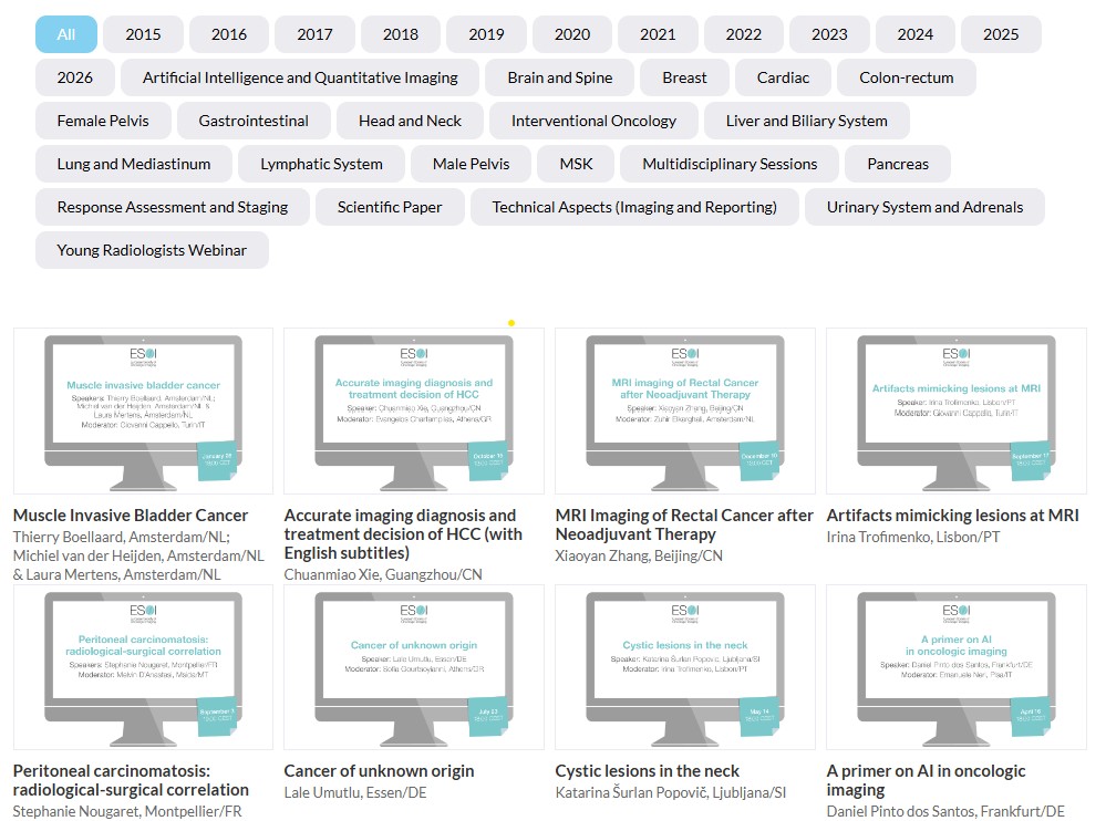
Find below comprehensive overviews of our previous webinar topics, year by year (2015-2025), to understand what we offer our active members in the form of recorded online lectures!
Previous Webinars 2025
Webinar #1: Lung cancer staging: changes, benefits and limitations
Sara Pilotto, Verona/IT & Helmut Prosch, Vienna/AT
Webinar #2: A multidisciplinary Approach to Diagnosis and Management of Patients with Endometrial Cancer
Niccolo Bizzarri & Silvia Bottazzi & Camilla Nero & Konstantinos Zormpas-Petridis; Rome/IT
Webinar #3: Primary bone tumors and mimickers
Salvatore Gitto, Milan/IT
Webinar #4: A primer on AI in oncologic imaging
Daniel Pinto dos Santos, Frankfurt/DE
Webinar #5: Cystic lesions in the neck
Katarina Šurlan Popović, Ljubljana/SI
Webinar #6: Peritoneal carcinomatosis: radiological-surgical correlation
Stephanie Nougaret, Montpellier/FR
Webinar #7: Cancer of Unknown Origin
Lale Umutlu, Essen/DE
Webinar #8: Artifacts mimicking lesions at MRI
Irina Trofimenko, Lisbon/PT
Webinar #9: Accurate imaging diagnosis and treatment decision of HCC
Chuanmiao Xie, Guangzhou/CN
Webinar #10: MRI Imaging of Rectal Cancer after Neoadjuvant Therapy
Xiaoyan Zhang, Beijing/CN
Previous Webinars 2024
Webinar #1: Cystic pancreatic neoplasms: MDT
Giuseppe Malleo, Verona/IT & Giulia Zamboni, Verona/IT
Special Webinar #1: Urothelial cancer
Davide Prezzi, London/UK
Webinar #2: Imaging of mediastinal masses
Helmut Prosch, Vienna/AT
Webinar #3: Common errors in oncologic imaging
Daniele Regge, Torino/IT
Webinar #4: Imaging after nonsurgical therapies for non-small cell lung cancer: A guide for radiologists
Marie-Pierre Revel, Paris/FR; Anna Rita Larici, Rome/IT
Webinar #5: GEP-NET: MDT
Massimo Falconi, Milan/IT; Francesco Panzuto, Rome/IT; Damiano Caruso, Rome/IT; Andrea Laghi, Rome/IT
Webinar #6: The 2021 WHO classification of CNS tumors: What the radiologist needs to know
Carolina Giordano, Rome/IT ; Simona Gaudino, Rome/IT
Webinar #7: Imaging considerations in appendicular soft tissue tumours
Christian Kelly-Morland, Framlingham/UK
Webinar #8: MDT discussion for cervical cancer
Benedetta Gui, Rome/IT; Luca Russo, Rome/IT
Webinar #9: Non-invasive imaging in the diagnosis and prognosis prediction of liver cancer
Yan Fuhua, Shanghai/CN
Previous Webinars 2023
Webinar #1: ESOI-ESSO-ESTRO: Rectal Cancer: A multi-disciplinary approach to imaging
Regina Beets-Tan, Geerard Beets; Amsterdam/NL
Webinar #2: Primary and secondary malignancies of the pleura: Diagnosis, staging and response assessment
Luciano Cardinale, Orbassano/IT
Webinar #3: Gastrointestinal cancers in IBD
Sofia Gourtsoyianni, Athens/GR
Webinar #4: Endometrial carcinoma
Milagros Otero-Garcia, Vigo/ES
Webinar #5: Whole-body diffusion weighted imaging
Vincent Vandecaveye, Leuven/BE
Special Webinar #1: Added value of artificial intelligence with CT and MRI for RCC diagnosis.
Zhen Li, Wuhan/CN
Special Webinar #2: Hepatospecific contrast agents in the study of HCC: Is the examination time the most important aspect?
Jin Wang, Guangzhou/CN
Webinar #6: Atypical presentations of bone mets
Frederic Lecouvet, Brussels/BE
Special Webinar #3: AI-powered Screening/Prognosis/Diagnosis: What’s the future of Oncology Imaging?
Luis Marti-Bonmati, Valencia/ES
Webinar #7: Rad-path correlations in breast cancer
Alexandra Athanasiou, Athens/GR
Webinar #8: Nuclear medicine for the diagnosis and treatment of neuroendocrine tumors
Clemens Cyran, Munich/DE
Webinar #9: Role of imaging in esophageal cancer
Vicky Goh, London/UK
Webinar #10: Imaging and staging of laryngeal cancer
Minerva Becker, Geneva/CH
Webinar #11: Update in interventional oncology of HCC
Laura Crocetti, Pisa/IT
Webinar #12: Characterisation of the Small Solid Renal Mass: When you need MRI
Davide Prezzi, London/UK
Special Webinar #4: Parametric MR before prostate biopsy, may increase patient life with PSA increased level
Tobias Franiel, Jena/DE
Previous Webinars 2022
Webinar #1: Multidisciplinary approach to small renal masses
Andrea Veltri, Orbassano/IT; Alessandro Volpe, Piedmont/IT; Marcello Tucci, Asti/IT
Webinar #2: O-Rads for MRI
Isabelle Thomassin-Naggara, Paris/FR
Webinar #3: Tumors and tumor-like lesions of the paranasal sinuses
Katarina Šurlan Popović, Ljubljana/SI
Webinar #4: Pediatric GI or GU tumors: A primer for the “adult radiologist”
Annemieke Littooij, Utrecht/NL; Irmina Sefic, Sarajevo/BA
Webinar #5: PET imaging evaluation in oncology: Tips & tricks for the clinical practice
André Dias, Aarhus/DK
Webinar #6: Cancer in pregnancy
Charis Bourgioti, Athens/GR
Webinar #7: Understanding Artificial Intelligence: Principles and basic concepts behind the “black box”
David Bonekamp, Hirschberg/DE
Webinar #8: Added value of PET/CT in staging and response assessment of head and neck tumors
Clemens Cyran, Munich/DE
Webinar #9: Abdominal toxicities of common oncologic therapies
Nina Tunariu, Tadworth/UK
Webinar #10: Incidental breast lesions at CT
Paola Clauser, Vienna/AT
Webinar #11: Life as an academic radiologist
Evis Sala, Cambridge/UK
Webinar #12: Skull base tumors: Differential diagnosis and key findings for treatment planning
Alexandra Borges, Lisbon/PT
Previous Webinars 2021
Webinar #1: ESOI-ESSO-ESTRO: Rectal Cancer: A multi-disciplinary approach to imaging
R. Beets-Tan (Radiologist), G. Beets (Surgeon), C. Marijnen (Radiation oncologist); Amsterdam/NL
Webinar #2: Imaging of lymphomas: Current status
M. Mayerhöfer, New York/US
Webinar #3: ESOI-ESHI-MT: AI in multimodality imaging of cancer
C. Cyran, Munich/DE
Webinar #4: Multidisciplinary webinar: Preoperative assessment of peritoneal carcinomatosis
S. Nougaret, F. Quenet; Montpellier/FR
Webinar #5: NET: How to assess response to therapy – A case-based multidisciplinary discussion
D. Caruso, M. Rinzivillo; Rome/IT (case presenters); F. Panzuto, Rome/IT; M. Falconi, Milan/IT (panelists)
Webinar #6: Preparing and attending oncologic MDT meetings: A memorandum for the radiologist
L. Fournier, Paris/FR
Webinar #7: ESOI-EUSOBI: Surveillance after breast cancer treatment
Kristina Lang, Lund/SE
Webinar #8: Urologic webinar:
– Penile cancer: Role of MRI in the new 8th TNM staging system
– Testicular germ cell tumors: Staging and surveillance
Alberto Vargas, Sungmin Woo, Anton Becker; New York/US
Webinar #9: ESOI-ESNR: Brain metastases: Imaging after treatment
Pia Sundgren, Lund/Sweden
Webinar #10: ESOI-ESTI: Pulmonary complications in oncologic therapy
What the oncologic radiologist must know
Cornelia Schaefer-Prokop, Amersfoort/NL
Webinar #11
Imaging of HCC:
– Radiomics and radiogenomics in HCC
– Assessment of HCC response to therapy: New treatments, new challenges, new criteria
Emanuele Neri, Riccardo Lencioni; Pisa/IT
Previous Webinars 2020
Limits and pitfalls of CT and MR in the staging of lung cancer
J. Dinkel, Munich/DE
Uterine tumours: Correlation between histology and imaging features
E. Sala (Radiologist), M. Jimenez-Linan (Pathologist); Cambridge/UK
A practical approach for the diagnosis of a liver tumor
V. Vilgrain, Paris/FR
Tumour board: GEP-NET
A. Laghi (Radiologist), D. Caruso (Radiologist), F. Panzuto (Gastroenterologist), M. Rinzivillo (Gastroenterologist); Rome/IT
M. Falconi, Milan/IT (Surgeon)
MSK – soft tissues tumours
F. Vanhoenacker, Antwerp/BE
Role of cardiac imaging (CT and MRI) in patients with cancer
B. Velthuis, Utrecht/NL
Spinal tumours
L. van den Hauwe, Antwerp/BE
Imaging biomarkers in oncology
A. Alberich-Bayarri, Valencia/ES
Multidisciplinary Webinar from ESOI-ESTRO
Lung cancer: A multidisciplinary approach to imaging
A.R. Larici (Radiologist), V. Valentini (Radiation Oncologist); Rome/IT
Oncologic Imaging and Covid-19: An unwanted wedding
D. Regge, Turin/IT; D. Caruso, Rome/IT
Imaging in gastric cancer before and after neoadjuvant therapy
M.A. Mazzei, Siena/IT
ESOI Young Radiologists’ Webinar: Basic statistics
M. Maas, Amsterdam/NL
Previous Webinars 2019
Prostate cancer: A multidisciplinary approach to imaging
H-P. Schlemmer (Radiologist), J.P. Radtke (Urologist); Heidelberg/DE
Immune therapy: Response assessment and toxicity
D. Regge, Turin/IT
Small bowel tumours
M. Gollub, New York/US
Interventional oncological options for RCC
M. Krokidis, Cambridge/UK
Rectal cancer: A multidisciplinary approach to imaging
R. Beets-Tan, Amsterdam/NL (Radiologist), N. Figueiredo, Lisbon/PT (Surgeon), R. Glynne-Jones, Northwood/UK (Radiation Oncologist)
Imaging and response assessment of multiple myeloma
A. Baur-Melnyk, Munich/DE
Artificial intelligence in oncologic imaging: Current status and future directions
E. Neri, Pisa/IT (Radiologist); A. Alberich Bayarri, Valencia/ES (Telecommunications Engineer)
Clinical relevance of PSMA PET/CT: An overview and update
C. Cyran, Munich/DE
Assessing response to therapy of brain tumours
P.M. Sundgren, Lund/SE
Pancreatic cancer: A multidisciplinary approach to imaging
G. Zamboni (Radiologist), G. Marchegiani (Surgeon); Verona/IT
Breast cancer: A multidisciplinary approach to imaging in neoadjuvant treatment
M. Lobbes, Sittard-Geleen/NL (Radiologist); M.L. Smidt, Maastricht/NL (Surgeon)
Interventional oncological treatment of lung nodules/lung cancer
J. Palussière, Bordeaux/FR
New paradigms in scientific publishing
M.F. Reiser, Munich/DE
Correlation of radiological images with surgical findings in ovarian peritoneal disease
L. Fournier (Radiologist), A.S. Bats (Surgeon); Paris/FR
Previous Webinars 2018
Rectal Cancer: A multidisciplinary approach to imaging
R. Beets-Tan (Radiologist), G. Beets (Surgeon), B. van Triest (Radiation oncologist); Amsterdam/NL
Cross-sectional imaging of renal tumours
R. Pozzi Mucelli, Verona/IT
Structured reporting in prostate cancer
D. Regge, Turin/IT
Imaging of testicular tumours
M. Bertolotto, Trieste/IT
Preoperative local staging of breast cancer: tailoring the surgical treatment
J. Camps Herrero, Alzira/ES (Radiologist); R. Ponzone, Turin/IT (Surgeon)
Response evaluation in oncology: RECIST 1.1
M. D’Anastasi, Msida/MT
Algorithmic approach to the female pelvic mass
E. Sala, Cambridge/UK
Molecular and hybrid imaging in prostate cancer
G. Cook, London/UK
Rectal cancer: A multidisciplinary approach to imaging
R. Beets-Tan (Radiologist), G. Beets (Surgeon), B. van Triest (Radiation oncologist); Amsterdam/NL
Diagnosis and staging of oesophageal and gastric cancer
M. Laniado, Dresden/DE
Neuroendocrine tumours – radionuclide imaging and therapy
H. Ilhan, Munich/DE
Structured reporting in rectal cancer
M. Maas, Amsterdam/NL
Imaging after treatment of lung cancer
J. Dinkel, Munich/DE
Multidisciplinary management of CRC liver metastases
Th. Helmberger, Munich/DE
Lung cancer screening – what did we learn so far?
C. Schaefer-Prokop, Amersfoort/NL
Assessment of the treatment response of bone lesions in cancer patients: Pearls and pitfalls
F. Lecouvet, Brussels/BE
Imaging and response assessment of gastrointestinal stromal tumours
A. Laghi, Rome/IT
Previous Webinars 2017
Imaging of CNS tumours
P. Parizel, Antwerp/BE
Imaging and differential diagnosis of orbital tumours
U. Mueller-Lisse, Munich/DE
Imaging of lymphomas
M. Mayerhöfer, Vienna/AT
Imaging spectrum of primary and secondary mesenteric neoplasms
S. Nougaret, Montpellier/FR
Imaging of the treated pelvis: Pearls and pitfalls
E. Sala, New York/US
MR imaging of malignant diseases of the uterus
Y. Lakhman, New York/US
Practical advice in preparing successful scientific manuscripts
M.F. Reiser, Munich/DE
Imaging of recurrent prostate cancer
H. Vargas, New York/US
Bone metastases, how should we approach them with imaging?
T. Bäuerle, Erlangen/DE
PET-MRI in oncologic imaging – where do we stand?
V. Goh, London/UK
When to call a lesion an HCC?
G. Brancatelli, Palermo/IT
Current evidence for minimally invasive treatments in HCC
J. Ricke, Munich/DE
Diagnostic test studies: How I do it
M. Maas, Amsterdam/NL
Paediatric abdominal tumours
C. Granata, Genoa/IT
Previous Webinars 2016
Staging of rectal cancer with MRI
R. Beets-Tan, Amsterdam/NL
Contrast-enhanced ultrasound-technical considerations and applications in oncologic imaging
D. Clevert, Munich/DE
Imaging dissemination pathways in head and neck cancer
M. Mack, Munich/DE
Whole body MRI in onology
V. Vandecaveye, Leuven/BE
Imaging of lung cancer: Implications on management
L. Bonomo, Rome/IT
Imaging of biliary tract tumours
R. Manfredi, Verona/IT
Staging of ovarian cancer – providing a GPS to patient management
H. Hricak, New York/US
Role of MRI in the diagnosis and management of prostate cancer
H. Vargas, New York/US
Tailoring breast imaging surveillance to the woman´s risk
F. Gilbert, Cambridge/UK
Imaging of bone tumours
G. Guglielmi, Foggia/IT
RECIST, CHOI and beyond
M. D’Anastasi, Munich/DE
Previous Webinars 2015
DW in oncologic imaging
D. Lambregts, Maastricht/NL
CT colonography
A. Laghi, Latina/IT
Cystic and solid pancreatic tumors
G. Zamboni, Verona/IT
Imaging the prostate – current status and future developments
J. Futterer, Nijmengen/NL
Update on breast MRI
F. Sardanelli, Milan/IT
Structured oncologic reporting
W. Sommer, Munich/DE
Webinar Recordings
Stay up-to-date in oncological imaging and enjoy unlimited access to a vast array of lectures from world leaders in their field!
ESOI webinars are recorded and made available to our active members (after a period of approximately 30 days).
To date, there are more than 100 full length recorded lectures available. In order to see which webinars have been organised in the past, please refer to the Webinar Overviews above. In addition, below is a preview of some lectures that are available.
In order to access the recordings via the link below, please make sure your membership 2026 is active and that you are logged in (members area – right upper corner).